uk-listings
Medical universities and training hospitals face continuous pressure to provide students with accurate, comprehensive anatomical education. Traditional methods rely heavily on physical specimens, which present logistical, financial, and ethical challenges. As technology advances, medical institutions are adopting advanced 3D modeling and digital simulation platforms to bridge the gap between theoretical knowledge and practical application. These digital systems provide a highly accurate, repeatable, and scalable solution for modern medical curriculums, standardizing the learning experience for future healthcare professionals.
Overcoming the Limitations of Physical Specimens
For decades, human cadavers have served as the primary resource for anatomical instruction. However, acquiring and maintaining these specimens requires specialized facilities, strict ethical compliance, and significant recurring costs. Furthermore, physical specimens degrade over time and allow for only a single dissection process. Once a tissue structure is altered or removed, the action cannot be undone.
This limits the number of students who can gain hands-on experience from a single specimen. Institutions now seek digital alternatives that offer repeatable, non-destructive exploration of human anatomy. This approach completely removes the storage and preservation requirements associated with formalin-fixed specimens.
Achieving Sub-Millimeter Data Accuracy
The transition to digital models requires uncompromising data accuracy. Systems developed from high-precision digital human datasets reconstruct human anatomy using original sectional data. These platforms extract voxels to build three-dimensional geometric models of organs, bones, muscles, blood vessels, and nerves.
High-end systems utilize datasets with a voxel size of 0.0384mm by 0.0384mm by 0.1mm. This level of granular detail ensures that the visual perception of the digital model matches that of a real anatomical specimen. Medical students can observe intricate nervous systems and micro-vascular structures that are often difficult to isolate in physical bodies.
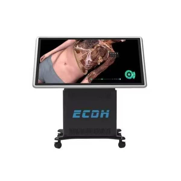
Through the implementation of a virtual anatomy table, educators can present these minute details on a large, interactive touch screen. This hardware allows multiple students to collaborate, dissect, and learn simultaneously. Digital platforms transform passive observation into active learning. Users can rotate, zoom, and dissect structures layer by layer.
If a student makes an error during a digital dissection, the system resets instantly. This provides a risk-free environment for surgical planning and anatomical exploration. The software includes features to isolate specific systems, such as the cardiovascular or skeletal system, hiding other tissues to provide an unobstructed view.
Synergy Between Digital Data and 3D Printing
The utility of these high-precision datasets extends beyond digital screens. The exact volumetric data used in digital platforms serves as the foundation for physical anatomical models. By utilizing full-color and multi-material 3D printers, institutions can create physical replicas of the digital structures. The 3D printing process extracts voxels from the surface of each anatomical structure within the original section dataset. This generates a texture map that ensures the printed geometric model looks visually identical to a real anatomical specimen.
Advanced 3D printers utilize 12 material channels to realize multi-material combined printing. Depending on the educational requirement, models can be printed using a combination of soft and hard materials, standard rigid materials, or even transparent packaging materials. These printed models possess a weight and tactile feedback similar to real tissue.
The authenticity and accuracy of these physical models earn recognition from anatomists, hand surgeons, and brain surgeons. This hybrid approach—combining digital interaction with tangible 3D printed models—provides a 1:1 simulation experience, particularly useful for institutions facing a severe lack of cadaveric specimens.
Immersive Learning with VR and AR Integration
To further enhance spatial understanding, modern educational providers integrate VR and AR technologies into their product ecosystems. Companies like DIGIHUMAN develop medical anatomy teaching products that allow students to explore three-dimensional structures in an immersive virtual environment.
By wearing a VR headset, a student can virtually walk through complex vascular networks or observe the spatial relationships of internal organs from an internal perspective. This level of immersion accelerates the learning curve for understanding complex spatial relationships, a fundamental requirement for accurate surgical navigation.
Financial and Operational Efficiency for Institutions
Budget considerations dictate educational resource procurement. Traditional gross anatomy labs require complex ventilation systems to manage chemical fumes, alongside the costs of waste disposal and continuous specimen procurement. Digital dissection platforms require an initial hardware and software investment but involve minimal recurring costs.
The software receives routine updates featuring new pathological cases and anatomical variations. This expands the institution’s library without requiring additional physical storage space. Over a standard hardware lifecycle, the cost per student decreases significantly compared to the continuous upkeep of a traditional wet lab.
Enhancing Communication in Clinical Environments
Beyond university education, high-fidelity digital models serve a functional purpose in clinical environments. Surgeons utilize 3D anatomical platforms for preoperative planning. By visualizing specific patient data or standard anatomical structures digitally, medical teams map out surgical approaches before entering the operating room. Additionally, these models assist in doctor-patient communication. Physicians use clear, three-dimensional visuals to explain complex diagnoses and proposed surgical procedures to patients. This visual aid improves patient comprehension and consent.
The integration of high-precision digital modeling into medical education and clinical practice represents a functional shift toward scalable, detailed, and repeatable learning. By utilizing advanced rendering techniques and highly accurate datasets, medical institutions provide a level of anatomical insight that meets the rigorous demands of modern healthcare training. The continuous development of these digital tools ensures that medical professionals receive comprehensive, accessible, and standardized education.